Cardiovascular Adaptations in Spinal
Cord-Injured Individuals

Patricia
de Groot, MsC, PT (Researcher)
Maria
Hopman, MD, PhD (Project
Leader)
Dirk
van Kuppevelt, MD![]()
On November 23, 2005, Patricia de Groot, defended her dissertation entitled:
Cardiovascular adaptations in spinal cord-injured individuals.
Summary
A spinal cord injury (SCI) disrupts the structural and functional integrity of the spinal cord and results in muscle paralysis, loss of sensation and autonomic dysfunction below the level of the injury. Although life expectancy in SCI has increased considerably the past decades, persons with SCI are still at increased risk for vascular related complications such as pressure ulcers, impaired wound healing and thromboses. In addition, well-known risk factors for cardiovascular diseases such as glucose intolerance, type 2 diabetes, disturbed lipid profile and alterations in body composition are all observed in higher proportion in the SCI population. Previous studies have shown that extensive central and peripheral circulatory changes occur in individuals with a chronic SCI. These vascular adaptations include a reduction in cardiac mass, reduced venous compliance, and. on the arterial side; a reduction in femoral artery size, decreased femoral compliance, reduced blood flow and almost doubled shear stress level in the femoral artery. Nowadays, the importance of physical inactivity as an independent risk factor for atherosclerosis and cardiovascular diseases is well recognized. Endothelial dysfunction plays an important role in the pathogeneses of cardiovascular diseases and impaired endothelium dependent dilation is directly linked with cardiovascular morbidity and mortality. Current knowledge on structural and function vascular adaptations that occur as a result of inactivity in humans is limited and dispersed. Most evidence on vascular changes, especially on the time course of vascular changes is derived from animal studies. The SCI population represents the lowest on the total spectrum of deconditioning in humans and offers a unique opportunity to assess the time course of vascular adaptations to inactivity and paralyses as well as to subsequent increased physical activity such as after functional electrical stimulation training. This thesis includes studies that relate to the topic cardiovascular adaptations in spinal cord-injured individuals. The main objective was to assess the magnitude and time course of arterial adaptations in vascular dimension and function after extreme inactivity in humans.
In the first part of
chapter 1, general information is provided about the spinal cord
population. An overview is presented on the known cardiovascular
adaptations in this patient population and we describe the potential to
(partly) reverse the detrimental vascular adaptations by means of functional
electrical stimulation training of the paralyzed muscles. Subsequently, a
physiological background is provided on the regulation of arteries. The blood
 flow
and diameter of arteries is the result of i) fast, functional changes
in vascular tone, and ii) arterial remodelling, which represents the
process of chronic and structural
changes in the arterial system. Furthermore, the current knowledge on the
magnitude and time course of functional and structural vascular adaptations to
inactivity is described from available data from different models of
deconditioning in animals (vascular ligation and hindlimb unloading) and
humans (bed rest,
spaceflight, limb immobilization and spinal cord injury). In addition,
information is provided on vascular adaptations following training with
emphasis on the time course of vascular changes. In summary, evidence from
animal studies indicates a rapid onset of vascular adaptations, i.e.
functional changes within days, followed by structural remodelling of the
vessel diameter within days to weeks. In humans, the current literature
derived from different models of deconditioning and exercise training studies
suggests that vascular adaptations to (in)activity may occur within weeks, but
the exact time course is not clear.
flow
and diameter of arteries is the result of i) fast, functional changes
in vascular tone, and ii) arterial remodelling, which represents the
process of chronic and structural
changes in the arterial system. Furthermore, the current knowledge on the
magnitude and time course of functional and structural vascular adaptations to
inactivity is described from available data from different models of
deconditioning in animals (vascular ligation and hindlimb unloading) and
humans (bed rest,
spaceflight, limb immobilization and spinal cord injury). In addition,
information is provided on vascular adaptations following training with
emphasis on the time course of vascular changes. In summary, evidence from
animal studies indicates a rapid onset of vascular adaptations, i.e.
functional changes within days, followed by structural remodelling of the
vessel diameter within days to weeks. In humans, the current literature
derived from different models of deconditioning and exercise training studies
suggests that vascular adaptations to (in)activity may occur within weeks, but
the exact time course is not clear.
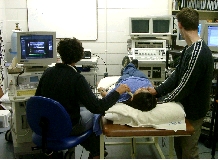
Classical venous occlusion plethysmography (VOP) of the leg, often used to assess venous compliance, measures properties of the whole calf, including volume changes at the arterial side and the interstitial fluid accumulation that occurs due to the enhanced capillary pressure during venous occlusion. Chapter 2 presents an ultrasound technique to measure the compliance of one major conduit vein in the leg. Ultrasound measurements of the popliteal vein were compared with classical VOP measurements, which were performed simultaneously in one subject. Short and long-term reproducibility of the measurements was assessed in a group of able-bodied subject and a comparison of venous compliance was made between healthy control subjects and individuals with known pathological changes in the venous vascular system (spinal cord-injured individuals). Results of the study demonstrate that the ultrasound and VOP measurements of venous compliance correlated significantly. Furthermore, we observed that ultrasound provides reproducible measurements with short and long-term coefficients of variation ranging from 10 to 15% for popliteal vein compliance and from 2 to 9% for absolute diameters at the different venous pressure steps. In addition, using ultrasound an 80% reduction in the compliance of the popliteal vein in SCI individuals compared with able-bodied controls was detected. We concluded that ultrasound is a suitable and reproducible method to measure conduit vein compliance. This technique provides the possibility to specifically assess compliance of one vein instead of the whole calf and appears to be a useful complementary method to the traditional venous occlusion plethysmography.
Changes in physical activity lead to marked changes in cardiac structure, ranging from the "physiological hypertrophy" of the endurance-trained athlete, to the "physiological atrophy" of chronically deconditioned patients. Although changes in cardiac dimensions with different modes of inactivity seem to be well described, the effect of inactivity on cardiac function has hardly been investigated. In Chapter 3, the effect of chronic deconditioning is assessed on cardiac morphology and function in a group of cervical spinal cord-injured individuals, who serve as human model of extreme inactivity. Echocardiographic measurements were performed to measure resting cardiac dimensions, (global and long-axis) diastolic and systolic function. The results demonstrate that tetraplegia is associated with a significant reduction in cardiac mass and left ventricular dimensions compared with able-bodied controls. However, resting diastolic and systolic function is not altered with continued exposure to inactivity, suggesting a remodelling of the heart as a physiological adaptive process.
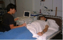
Animal experiments have shown that vascular adaptations occur within days/weeks. However, the time course of vascular adaptations to inactivity in humans is not clear. Folkow hypothesized that structural vascular adaptations in humans may be largely completed within a few months, presumably reflecting the five to six fold lower metabolic rate in humans than in small animals such as rats and rabbits. In Chapter 4, the time course of vascular adaptations from 6 weeks to 13 months after a spinal cord injury is studied. Arterial diameters and red blood cell velocity of the common femoral artery, carotid artery and brachial artery were measured using echo Doppler ultrasound. In a cross-sectional study design, a group of SCI individuals with time since lesion varying from 6 weeks to 13 months post-injury were measured on one occasion. In addition, longitudinal measurements were performed at week 6, 8, 12, 16, 20 and 24 after the trauma in a group of 6 SCI individuals. Results show that within 6 weeks after a spinal cord injury, diameter (-30%) and blood flow (-30%) are significantly reduced and peak (+50%) and mean wall shear rate (+100%) of the femoral artery are significantly increased. No further changes in femoral arterial properties were observed between week 6 and 13 months post-injury. Carotid and brachial arterial properties were not different from controls and did not change over time. Thus, the process of vascular adaptations to inactivity and paralyses in humans seems to be largely completed within six weeks.
In chapter 5 we studied the time course of adaptations in vascular dimension and function during the first six weeks after a spinal cord injury. Six male spinal cord-injured patients were included immediately after their injury and vascular dimension, blood flow, endothelial function and limb volume were measured at 1, 2, 3, 4 and 6 weeks post-injury. Vascular characteristics of the common femoral artery, superficial femoral artery, brachial artery and carotid artery were assessed with echo Doppler ultrasound. Endothelial function in superficial femoral artery was measured with Flow Mediated Dilation (FMD). A rapid onset of vascular adaptations in the inactive and paralyzed legs after acute spinal cord injury was observed. Changes include an approximately 25% reduction in femoral artery size and leg volume, a doubling in basal shear rate levels and a significant increase in FMD responses. All these adaptations are largely accomplished within 3 weeks post- injury.
Impaired endothelial function has been suggested as a key early event in the development of atherosclerosis and a high correlation between endothelial dysfunction and risk factors for cardiovascular diseases including hypertension, hypercholesterolemia, cigarette smoking, diabetes and ageing has been reported. Besides above-mentioned traditional risk factors, it is well-known that physical inactivity is associated with an increased risk of developing cardiovascular diseases. However, at present, the relationship between inactivity and endothelial function is not clear. In individuals with paraplegia, the part of the body below the lesion level is paralyzed and thus extremely inactive. In contrast, the upper limbs are often relatively active since the arms are used for ambulation due to their wheelchair bound life style. In chapter 6, conduit artery endothelial function, measured by Flow Mediated Dilation (FMD), in an inactive extremity (leg, superficial femoral artery) and chronically active extremity (arm, brachial artery) within one subject was studied. Eleven male SCI individuals and eleven male able-bodied controls were included. Echo Doppler measurements were performed to measure FMD responses after 10 and 5 minutes of arterial occlusion of the leg and the arm, respectively. A nitroglycerine spray was administered to determine the endothelium independent vasodilatation in the superficial femoral artery. The results demonstrate that vascular endothelial function, expressed as percentage change in FMD; is enhanced in the femoral artery of the SCI individual compared with controls whereas no differences between the groups were found in the relative FMD response of the brachial artery. When taken the stimulus into account (using the ratio of FMD/delta shear rate as index for endothelial function) the results indicate that spinal cord-injured individuals have at least a preserved endothelial function in the inactive legs and possibly an attenuated endothelial function in the active arms as compared with able-bodied controls.
Both endothelial dysfunction
and decreased arterial compliance have been shown to be associated with the
incidence and progression of cardiovascular diseases. Regular endurance
exercise has been shown to improve endothelial function and previous
cross-sectional studies have demonstrated that compliance in the femoral
artery is significantly higher in endurance trained athletes compared with
sedentary controls and SCI individuals.
It was previously
reported that relative FMD response was increased in the inactive legs of SCI
individuals compared with control subjects (chapter 5). The finding of an
enhanced FMD response in
 deconditioned
arteries may be surprising since it has been shown previously that FMD
increases following exercise training. It is not known whether the changes in
arterial compliance and endothelial function in SCI are reversible by training
after a prolonged period of extreme deconditioning and what the time course of
these training induced vascular adaptations might be. Animal research and
recent human evidence suggest that vascular adaptations occur within days or
weeks of the onset of training. In chapter 7 we studied the effect and
the time course of enhanced physical activity after a long period of
deconditioning on central and peripheral arterial compliance and on
endothelial function. Arterial compliance was measured in the superficial
femoral, brachial and carotid arteries and local FMD responses were measured
in the superficial femoral and brachial artery prior to, and after I, 2, and 4
weeks of 30 minutes of daily leg functional electrical stimulated (FES)
training in a group SCI individuals. The training included one leg only, while
the other leg was used as a time control. The results of the study demonstrate
that daily electrically induced training of an extremely deconditioned leg in
SCI individuals normalizes their initially enhanced FMD response (within 2
weeks) and elevates arterial compliance and hyperemic flow (within 4 weeks) in
the femoral artery of the trained leg only. No changes over time were observed
in the vascular properties of the femoral artery of the untrained leg and the
brachial and carotid arteries. This encouraging conclusion may have important
implications for future clinical applications of electrical stimulation.
deconditioned
arteries may be surprising since it has been shown previously that FMD
increases following exercise training. It is not known whether the changes in
arterial compliance and endothelial function in SCI are reversible by training
after a prolonged period of extreme deconditioning and what the time course of
these training induced vascular adaptations might be. Animal research and
recent human evidence suggest that vascular adaptations occur within days or
weeks of the onset of training. In chapter 7 we studied the effect and
the time course of enhanced physical activity after a long period of
deconditioning on central and peripheral arterial compliance and on
endothelial function. Arterial compliance was measured in the superficial
femoral, brachial and carotid arteries and local FMD responses were measured
in the superficial femoral and brachial artery prior to, and after I, 2, and 4
weeks of 30 minutes of daily leg functional electrical stimulated (FES)
training in a group SCI individuals. The training included one leg only, while
the other leg was used as a time control. The results of the study demonstrate
that daily electrically induced training of an extremely deconditioned leg in
SCI individuals normalizes their initially enhanced FMD response (within 2
weeks) and elevates arterial compliance and hyperemic flow (within 4 weeks) in
the femoral artery of the trained leg only. No changes over time were observed
in the vascular properties of the femoral artery of the untrained leg and the
brachial and carotid arteries. This encouraging conclusion may have important
implications for future clinical applications of electrical stimulation.
Persons with a spinal cord injury are at increased risk for secondary complications such as pressure ulcers and attenuated wound healing because of an impaired circulation and concomitant decreased tissue perfusion. It has been suggested that passive exercise enhances the blood flow via mechanical pump effects or reflex activation. Passive leg movements carried out by a physical therapist and passive cycling are two forms of passive exercise, frequently used in rehabilitation of persons with SCI. Whether or not these interventions positively affect the arterial peripheral circulation is unknown. Therefore, the main objective of the study in chapter 8 was to assess peripheral circulatory responses during and after passive leg movements and passive cycling in individuals with SCI and able-bodied controls, using protocols in accordance with the clinical setting in rehabilitation centres. Echo Doppler measurements were performed to measure leg blood flow at rest, during and after ten minutes of standardised passive leg movements and twenty minutes of passive leg cycling, respectively. Blood pressure was measured continuously and total and leg vascular resistance was calculated. In both groups, no changes in leg blood flow, vascular resistance nor in blood pressure were observed during or after the two interventions. The results of the present study demonstrate that passive leg movements and passive cycling do not alter the arterial peripheral circulation in persons with SCI and controls. Although the present study does not support the use of passive movements or- exercise for prevention of cardiovascular related secondary complications, physical therapists should not be dissuaded from using this modality to address musculoskeletal concerns.
In the general discussion in chapter 9, the current knowledge on the time course and magnitude of arterial vascular changes to inactivity is reviewed from available data from human models of deconditioning with the emphasis on recent studies in this field by the authors, i.e. the effects on vascular dimension and function after 4 weeks of unilateral limb suspension, 52 days of strict horizontal bed rest (the Berlin Bed Rest Study) and the vascular changes that occur (within the first year) after a spinal cord injury (chapter 4, 5 and chapter 6 of this thesis). We conclude that in accordance with data derived from animal research, a rapid onset of vascular adaptations after inactivity is evident in humans. A substantial reduction in femoral diameter size was observed after I week of leg casting (6%),4 weeks of limb suspension (12%),8 weeks of bed rest (17%), and, in the most extreme on the spectrum of inactivity, a 25% reduction in diameter size was completed within only 3 weeks after a spinal cord injury. The human arterial system strives to maintain a constant shear stress by adapting the diameter to chronic changes in blood flow. Recent training studies in humans have suggested that conduit arteries adapt their baseline diameter to peak shear stress and peak oxygen consumption rather than to resting blood flow. Parallel to this reasoning, the decrease in diameter after deconditioning most likely represents an inward remodelling as an adaptation to a total lack of variation in peak shear stress and not to basal shear stress levels. During six weeks of extreme inactivity, a simultaneous decrease over time was observed for femoral artery size and limb volume, which suggests a strong functional link between adaptations in vascular dimension and muscular atrophy. In addition, decreased hyperaernic leg blood flow, an almost doubling of resting shear rate levels in the femoral artery and increased FMD responses in the superficial femoral artery were all are evident within 3 to 8 weeks in different humans models of inactivity. Possible explanations for an increased FMD response in deconditioned arteries may be related to the chronically enhanced baseline shear stress levels in leg conduit arteries. Since it well-known that shear stress is a potent physiological stimulus for NO release, the increased levels of basal shear stress may lead to an upregulation of eNOS. A second possible explanation may be the lack of periods of high shear stress in the deconditioned vessels, which may contribute to an up-regulation of NO responsiveness. The enhanced FMD responses in deconditioned arteries imply that the functional vascular adaptations to inactivity are not simply the inverse of adaptations to exercise.
Implications of the studies and future directions
The studies in the present thesis mainly focus on the magnitude and time course of changes in vascular dimension and function after a spinal cord injury, which obviously represents the most extreme on the total spectrum of deconditioning in humans. In order to apply effective interventions, insight into the time course of vascular adaptations is essential. The results of this thesis may have important clinical implications for the SCI population and complement current human vascular physiology knowledge on the magnitude and time course of vascular adaptations to extreme inactivity. We have shown that, as in animals, function and structural vascular changes in conduit arteries occur within weeks. Recently, it has been suggested that the mechanism and time course of vascular adaptations to exercise or inactivity may differ between distinct vascular beds, i.e. conduit and resistance arteries. This hypotheses however, needs further investigation and represent an interesting and relatively new field. Increased oxidative stress is associated with endothelial dysfunction and has been reported in several cardiovascular conditions including hypertension, arteriosclerosis and diabetes. It is known that changes in NO bioavailability occur to changes in the balance between free radical formation and its disposal by antioxidants. At present, the effect of deconditioning on oxidative stress and its impact on vascular function is unknown and future studies should be directed to assess the underlying mechanisms of deconditioning and conditioning on oxidative stress and vascular function.
Publications
from this thesis
Ultrasound: a reproducible method to measure conduit vein compliance. De Groot PC, Bleeker MW, Hopman MT. J Appl Physiol. 98(5): 1878-1883, 2005.
-
Time course of Arterial Vascular Adaptations to Inactivity and Paralyses in humans. De Groot PCE, HJM van Kuppevelt, C. Pons, G. Snoek, LHV van der Woude, MTE Hopman. Medicine and Science in Sports and Exercise, 35: 1977-1985, 2003.
Rapid and extensive arterial adaptations after spinal cord injury. De Groot PCE, Bleeker MWP, van Kuppevelt DHM, van der Woude LHV, Hopman MTE. Arch Phys Med Rehab 87(5): 688-696, 2006.
Preserved Flow-Mediated Dilation in the inactive legs of spinal cord-injured individuals. De Groot P CE, F Poelkens, M Kooijman, MTE Hopman. American Journal of Physiology Heart Circulatory Physiology. 287: H374-H380, 2004.
Electrical stimulation alters FMD and arterial compliance in extreme inactive
legs. De Groot PCE, Crozier J, Rakobowchuk M, Hopman MTE. Medicine
and Science in Sports and Exercise, 37:1356-1364, 2005.
Passive leg movements and passive cycling do not alter arterial leg blood flow in individuals with spinal cord injury. Ter Woerds W, De Groot PCE, Van Kuppevelt DHJM, Hopman MTE. Physical Therapy, 86(5): 636-645, 2006.
Magnitude and time course of arterial vascular adaptations to inactivity in
humans. De Groot PC,
Bleeker MWP, Hopman MTE. Exercise and Sport Sciences Reviews. 34(2):
65-71, 2005. Contact
address
Sonja
de Groot / Lucas van der Woude Center for Human Movement Sciences University Medical Center Groningen University of Groningen Antonius Deusinglaan 1 9713AV Groningen, The Netherlands
![]()